Key Takeaways
Effective intracellular delivery depends on more than cell entry; therapeutic cargo must reach the correct intracellular compartment in an active form.
Endosomal escape remains a major bottleneck for RNA therapeutics, macromolecular biologics, and other advanced modalities.
Cytosolic delivery assays, such as SLEEQ and Gal8 recruitment imaging, can help distinguish productive cargo release from simple cellular uptake.
Nuclear targeting adds another layer of complexity for genome-editing systems, requiring strategies that support cytosolic access, nuclear localization, and functional activity.
Cell Entry Is Only the Beginning
For many therapies, reaching the right tissue is no longer enough. The expanding universe of RNA therapeutics, protein and peptide medicines, macromolecular biologics, and genome-editing systems has shifted attention from the cell surface to the cell interior. These modalities often depend on access to intracellular sites of action, including the cytosol and the nucleus. A drug that binds a receptor or accumulates near a target cell may still fail if its active cargo cannot cross the plasma membrane, escape intracellular sequestration, and arrive intact at the compartment where its biological effect is required.
That distinction changes how delivery must be understood. Cellular uptake can create the appearance of success, because a carrier or cargo may be visible inside the cell. Yet uptake is not equivalent to functional delivery. Many platforms enter cells through endocytic pathways, which can place their cargo inside membrane-bound compartments rather than in the cytosol. From there, the cargo may be sorted, recycled, trapped, or degraded before it reaches its intended site of action. For intracellular medicines, the most important question is therefore not simply whether material enters a cell but whether enough active cargo reaches the right intracellular compartment to produce a meaningful biological response.
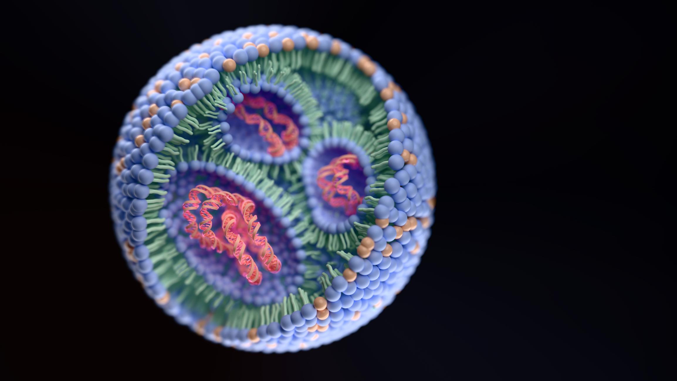
RNA therapeutics illustrate the challenge clearly. RNA molecules must overcome cellular barriers before they can realize their therapeutic potential, including the need to cross cellular membranes and avoid defenses that limit intracellular access. Lipid nanoparticles (LNPs) have helped bring RNA delivery into clinical use, including LNP–mRNA COVID-19 vaccines, but LNP delivery remains constrained by tissue tropism and delivery efficiency. Those limitations do not diminish the importance of LNPs, but they clarify why the next stage of delivery innovation will depend on understanding the intracellular journey in greater detail.
Macromolecular biologic therapeutics face similar barriers. Peptides, proteins, and small interfering RNAs (siRNAs) may require delivery agents to reach the cytoplasm or nucleus, and cell-penetrating peptides (CPPs) or protein transduction domains (PTDs) can promote uptake through endocytosis. However, endosomal escape remains a major challenge after that uptake has occurred. The same general lesson applies across multiple cargo classes: internalization is only the first step in a longer delivery pathway.
Here, we explore three linked dimensions of intracellular delivery: endosomal escape, cytosolic delivery, and nuclear targeting. Each represents a different point in the intracellular route, and each requires distinct design and measurement strategies. Endosomal escape determines whether cargo can leave endocytic compartments. Cytosolic delivery determines whether cargo becomes bioavailable in the compartment where many RNA, protein, and peptide modalities act. Nuclear targeting adds another barrier for systems that must reach DNA or other nuclear machinery. Treating these as separate but connected problems can help developers avoid optimizing the wrong endpoint.
The Endosome as a Delivery Checkpoint
Most intracellular delivery systems must contend with the endosome. Endocytic uptake provides a route into the cell, but it also places cargo within a membrane-bound pathway that is not designed to release therapeutics into the cytosol. Internalized material can remain sequestered in endosomes and ultimately be degraded in lysosomes, creating what is often described as the endosomal escape problem.1 In practical development terms, this means that a delivery vehicle can perform well in an uptake assay while still performing poorly as a therapeutic delivery system.
Endosomal escape is not a single formulation feature or a binary event. Endosomes mature, sort their contents, interact with repair pathways, and direct cargo toward different fates. A carrier that disrupts an endosomal membrane may still fail to release its payload efficiently. Cargo that separates from its carrier at the wrong time may be left behind, diverted, or degraded. These biological details matter because intracellular delivery depends on the relationship between the carrier, the cargo, and the cellular trafficking pathway.
LNP-mediated RNA delivery provides a useful example. Limited escape from endosomes into the cytosol is generally considered the rate-limiting step for RNA delivery.2 Several barriers can limit productive cytosolic delivery, including incomplete RNA release from damaged endosomes and segregation of RNA payload from ionizable lipid during endosomal sorting. Those mechanisms help explain why optimizing nanoparticle composition alone may not fully solve delivery efficiency. The system must support uptake, endosomal membrane interaction, cargo release, and productive diffusion of the cargo into the cytosol.
This has implications for how delivery platforms are designed. If endosomal escape is treated only as a membrane disruption problem, the result may be a carrier that damages endosomes without delivering enough active cargo. If it is treated only as a chemistry problem, teams may miss the importance of intracellular trafficking, cargo–vehicle separation, and biological repair mechanisms. The more precise view is that endosomal escape is a checkpoint at which formulation, cargo properties, cell biology, and timing intersect.
The importance of that checkpoint extends beyond RNA. Macromolecular biologics that enter through endocytosis face the same risk of endosomal entrapment. CPPs and PTDs can increase uptake, but uptake alone does not guarantee release into the cytoplasm.3 This distinction is especially important when total cellular association is used as an early screen. A molecule that associates strongly with cells may look promising, but that signal may not represent the fraction of cargo that becomes accessible to intracellular targets.
For development teams, the endosome should therefore be viewed less as an obstacle to be mentioned generically and more as a design environment. The relevant question is not only how to promote uptake but how to control the fate of cargo after uptake. That requires assays that can distinguish endosomal localization, membrane disruption, cytosolic release, and downstream biological activity.
Cytosolic Bioavailability, Not Just Cellular Uptake
For many intracellular modalities, the cytosol is the functional destination. Messenger RNA (mRNA) must reach the cytosol to be translated. siRNA must reach the cytosol to engage RNA interference machinery. Many therapeutic proteins and peptides also need cytosolic access to interact with intracellular targets. Cytosolic transport is therefore essential for therapeutic peptides, proteins, and nucleic acids, but it remains a major obstacle.4
The concept of cytosolic bioavailability helps sharpen the delivery challenge. A cargo may be present somewhere inside a cell, but only the fraction that reaches the cytosol in an active form can contribute to the intended effect. Total intracellular signal, total cell association, or uptake into vesicular compartments may overstate productive delivery. This problem is not merely semantic; it can influence platform selection, formulation optimization, and candidate progression.
Cell-penetrating peptides provide an instructive case. Positively charged CPPs can increase total cytosolic accumulation, yet that increase may reflect stronger nonspecific cell association rather than improved endosomal escape efficiency.4 In other words, a delivery strategy can increase how much cargo appears to get into cells without improving the step that matters most for release from endosomes. That finding reinforces the need to separate uptake, association, and escape as distinct performance measures.
Synthetic endosomal escape domains represent another approach to the same problem. Studies of engineered escape domains have investigated ways to enhance cytoplasmic delivery of macromolecular cargo, reflecting the broader need for delivery agents that can move biologic payloads beyond endosomal compartments.3 These strategies are part of a wider effort to convert endocytic entry into functional cytosolic access.
Nevertheless, cytosolic delivery cannot be reduced to a single universal mechanism. Different cargos impose different constraints. RNA molecules, proteins, peptides, and ribonucleoprotein complexes vary in size, charge, stability, structure, and intracellular sensitivity. A delivery system optimized for one cargo may not translate directly to another. The relevant success criteria also differ by mechanism of action. For mRNA, delivery must support translation. For siRNA, it must support loading into the appropriate RNA interference pathway. For proteins or peptides, it must preserve structural or binding activity. For genome-editing ribonucleoproteins, cytosolic access may be necessary but not sufficient if the system must ultimately enter the nucleus.
Cytosolic delivery is best understood as an intermediate functional endpoint rather than a final guarantee of efficacy. It marks the point at which cargo has escaped the endocytic pathway and entered a compartment where many intracellular mechanisms become possible. However, the cargo must still remain intact, available, and capable of engaging its biological target. A useful intracellular delivery strategy should therefore be evaluated across multiple layers: uptake, endosomal escape, cytosolic bioavailability, and mechanism-specific activity.
Assays That Separate Uptake from Escape
The development of intracellular delivery systems has been constrained by the difficulty of measuring the right events. Older or indirect methods can report uptake, total intracellular signal, or downstream activity without clearly identifying how much cargo actually escaped the endosome. This has contributed to conflicting interpretations of cytosolic delivery mechanisms and limited mechanistic understanding.4
More sensitive and more direct assays are beginning to change the conversation. One example is the split luciferase endosomal escape quantification assay, or SLEEQ, which was developed to probe cytosolic delivery and endosomal escape with high sensitivity.4 In the reported study, SLEEQ detected low levels of cytosolic delivery and measured endosomal escape efficiencies of approximately 2% in HEK293 cells and approximately 7% in HeLa cells for green fluorescent protein (GFP) under the tested conditions. Those values are useful not because they establish a universal benchmark but because they show how limited productive escape can be even when delivery appears measurable.
SLEEQ also highlights why assay design matters. By helping distinguish total cytosolic accumulation from endosomal escape efficiency, it can reveal whether a delivery strategy improves the escape step itself or simply increases nonspecific cell association.4 That distinction could be critical during formulation screening. A platform that appears superior in an uptake assay may not be superior when assessed for productive cytosolic release.
Galectin-based imaging provides another way to evaluate endosomal disruption. Galectin-8 (Gal8) recruitment imaging has been studied as a method to predict intracellular bioavailability of carrier formulations.5 In that work, Gal8 recruitment imaging was compared with other methods, including pH-dependent hemolysis profiling, LysoTracker colocalization, and total cellular internalization measurements. The approach has also been presented as a possible screening component for libraries of endosome-disrupting polymers and lipids.
These analytical methods support a broader shift in how delivery systems should be evaluated. Rather than asking only how much cargo is associated with cells, teams can begin asking whether the carrier damages endosomal membranes, whether the cargo escapes, whether the cargo becomes bioavailable in the cytosol, and whether that bioavailability produces the expected biological function. No single assay can answer all of those questions. A stronger development strategy would use complementary assays to connect physical delivery events with functional outcomes.
This matters for translation. When early screening relies on simplified proxies, candidates may be advanced because they perform well against a convenient measurement rather than because they solve the delivery problem that will determine efficacy. Conversely, a more mechanistic assay package may help explain why a platform fails, why it works only in certain cell types, or why a formulation change improves one parameter while worsening another. For intracellular delivery, analytics are not merely confirmatory but part of the design process.
What LNPs Have Taught the Field
LNPs are the most visible example of intracellular delivery reaching broad clinical relevance. Their use in mRNA delivery, including LNP–mRNA COVID-19 vaccines, demonstrated that nanoparticle-enabled RNA delivery could move from a specialized research area into the center of therapeutic development.6 That success has made LNPs a reference point for many discussions of intracellular delivery, even when the cargo, route, tissue target, or intracellular destination differs.
Their success has also clarified the limits of current delivery systems. LNPs remain constrained by tissue tropism and delivery efficiency, and endosomal escape remains a major bottleneck for RNA delivery.2 These limitations matter because LNP performance depends on more than particle formation or cellular uptake. The carrier must navigate biological barriers, interact with endosomal membranes, release RNA, and allow the RNA to diffuse productively within the cytosol.
The design space for LNPs has often focused on lipid chemistry, including ionizable lipids and helper components. That remains important, but emerging work suggests that internal nanoparticle architecture may also influence delivery performance. A 2026 study of engineered gold-core LNPs reported a twofold increase in endosomal escape and approximately 100-fold greater cytoplasmic mRNA diffusion compared with conventional LNPs under the conditions tested.7 The study’s authors framed internal LNP architecture as a design lever for overcoming the endosomal escape bottleneck.
That example should be interpreted carefully. It does not mean that gold-core LNPs have become a general solution, nor does it replace the need to evaluate safety, manufacturability, scalability, and clinical translation. It does, however, reinforce an important principle: delivery performance may depend on structural features that influence how cargo behaves after endosomal disruption. If endosomal damage occurs but RNA remains poorly released, poorly separated from the carrier, or poorly diffused into the cytosol, then uptake and membrane interaction will not translate into efficient expression.
The LNP field also shows why development considerations cannot be separated from delivery biology. Clinical translation of LNP–mRNA formulations requires attention to good manufacturing practice (GMP), stability, storage, and safety.6 Those considerations are not downstream administrative details. They shape which delivery designs can be developed, manufactured, characterized, stored, distributed, and used reliably.
For drug developers and development and manufacturing partners, the lesson from LNPs is not that every intracellular delivery problem should be solved with an LNP. It is that a successful delivery platform must be evaluated across biology, formulation design, analytical characterization, and translational feasibility. LNPs have provided proof that intracellular delivery can be clinically powerful. They have also shown why the next generation of platforms must be judged by what happens after cellular entry.
When The Target Is in the Nucleus
Cytosolic delivery is a major achievement for many modalities, but some therapeutic strategies require a further step: nuclear access. Genome editing provides the clearest example. A system that must act on DNA cannot stop at the cytosol. It must reach the nucleus in an active form, which adds another barrier after cellular uptake and endosomal escape.
Nuclear delivery requires navigation through several sequential obstacles. A nuclear-directed delivery system must cross the cell membrane, avoid endo/lysosomal entrapment and degradation, traffic through the cytoplasm, and enter the nucleus.8 These steps overlap with cytosolic delivery but are not identical to it. A formulation that successfully releases cargo into the cytosol may still fail if the cargo is too large, unstable, poorly localized, or unable to engage nuclear import mechanisms.
The nuclear pore complex (NPC) provides the main gateway between the cytoplasm and the nucleus. NPCs mediate most communication between these compartments, and small cargoes can diffuse through them while larger molecules generally require facilitated transport.9 This creates a size- and mechanism-dependent challenge for delivery systems. Nuclear targeting must account not only for release into the cytosol but also for transport across the nuclear envelope.
Nuclear localization signals (NLSs) are one widely used strategy for directing material to the nucleus. NLSs have been attached to nanoparticles in many applications to promote nuclear delivery.8 In principle, this approach uses existing cellular transport machinery to help move cargo toward or into the nucleus. In practice, however, the performance of NLS-based systems depends on cargo properties, carrier design, intracellular trafficking, and whether the NLS remains accessible and functional at the right point in the delivery pathway.
Nuclear targeting also raises analytical challenges. Measuring cytosolic escape may not be enough. A nuclear-directed system may require assays for endosomal escape, cytosolic availability, nuclear localization, and target engagement. It may also require a clearer understanding of whether the delivery vehicle itself enters the nucleus, whether the cargo separates from the vehicle before nuclear import, or whether only a subcomponent reaches the nuclear compartment. These questions can affect both efficacy and safety, particularly for systems that alter genetic material.
For development teams, nuclear targeting should be treated as a distinct design objective rather than an extension of uptake. The route to the nucleus includes the same early delivery challenges faced by cytosolic modalities, but it adds a second gate that has its own biological constraints.
CRISPR Shows Why Nuclear Delivery Matters
CRISPR/Cas9 systems offer a practical example of how intracellular delivery can require multiple sequential solutions. A ribonucleoprotein (RNP) system must enter the target cell, avoid degradation, become available intracellularly, and, when editing genomic DNA, reach the nucleus. Modified LNPs have been engineered for systemic delivery of CRISPR-Cas9 RNPs into cells and tissue editing in preclinical contexts.10 That work illustrates the broader interest in delivery systems that can handle large, complex biologic cargoes.
The nuclear step remains important. Enhancing intracellular delivery and performance of RNA-guided CRISPR-Cas9 nucleases remains an active need, and nuclear translocation of commonly used Streptococcus pyogenes Cas9 (SpCas9) proteins can be suboptimal.11 This means that delivery strategy cannot end with cell entry or even cytosolic release. The editing machinery must be positioned in the compartment where it can act.
NLS engineering is a common way to address this issue. Incorporating NLS sequences at one or both termini of CRISPR enzymes is widely used to facilitate genome editing.12 However, design choices can create tradeoffs. Terminal NLS fusion can negatively affect recombinant protein yield, which shows how a nuclear-targeting strategy can intersect with manufacturability. That is an important development lesson: a modification that improves intracellular localization may complicate expression, production, or characterization.
Other approaches are also under investigation. Non-NLS strategies for nuclear transport of Cas9/sgRNA RNPs have been explored, including systems using apoferritin nanocages and doxorubicin for nuclear transport of NLS-free Cas9/sgRNA RNPs.13 This does not establish a preferred route, but it shows that nuclear delivery remains an active design problem rather than a solved feature of genome-editing systems.
CRISPR also highlights the importance of matching the delivery endpoint to the therapeutic mechanism. For some cargoes, cytosolic bioavailability is sufficient. For CRISPR systems acting on genomic DNA, cytosolic access is only one stage. The delivery system must support nuclear localization, target engagement, and functional editing. Each stage introduces potential loss, variability, and design tradeoffs.
The development implications are significant. A CRISPR delivery system could fail because of poor tissue targeting, inefficient cellular uptake, endosomal entrapment, weak cytosolic release, inadequate nuclear translocation, insufficient editing, or manufacturing constraints related to the cargo or delivery format. Improving one stage may not improve the whole system if another stage becomes limiting. This is why intracellular delivery should be developed as an integrated pathway rather than a collection of isolated formulation attributes.
Designing Delivery Around the True Site of Action
Intracellular delivery strategy should begin with the site of action. A cytosolic RNA therapy, a cytosolic protein therapeutic, and a nuclear genome-editing system do not require the same delivery endpoint. They may share early barriers, including uptake and endosomal escape, but their success criteria diverge once cargo enters the cell.
This has practical consequences for formulation development. A carrier and cargo should be evaluated together because their intracellular fates may differ. In LNP-mediated RNA delivery, RNA payload and ionizable lipid can segregate during endosomal sorting, which may limit productive release even when endosomal disruption occurs.2 That finding supports a more integrated view of delivery, in which the behavior of the formulation after uptake matters as much as its properties before administration.
Assay selection should follow the same logic. Uptake, endosomal disruption, cytosolic bioavailability, nuclear localization, and functional activity are related but not interchangeable. A development program focused on intracellular delivery should avoid relying on a single proxy endpoint. Instead, it should build an analytical package that reflects the intended mechanism and identifies which step is limiting. Gal8 recruitment imaging, SLEEQ, localization studies, and functional assays can each contribute different information, but their value depends on how clearly they are connected to the therapeutic objective.4,5
The target cell type also matters. Delivery performance can vary across cellular contexts, and endosomal escape efficiencies measured in one model may not translate directly to another. The SLEEQ study’s reported efficiencies differed between HEK293 and HeLa cells under the tested conditions, which underscores the need to interpret assay results in context rather than as universal constants.4 For development teams, this means that relevant cell models, mechanism-appropriate assays, and context-specific interpretation are all essential.
Manufacturability should be considered early. The LNP–mRNA field has made clear that clinical translation requires GMP production, stability, storage, and safety considerations.6 CRISPR nuclear localization strategies also show that molecular engineering choices can affect recombinant expression yield.12 These examples point to the same development principle: delivery design must be compatible with production, characterization, and use at the intended stage of development.
This is especially relevant for partnerships among biopharma companies, formulation specialists, analytical teams, and manufacturing organizations. Intracellular delivery problems often sit at the boundaries of biology, chemistry, process development, and analytics. A carrier may look promising from a formulation perspective but fail to produce sufficient intracellular bioavailability. A cargo modification may improve localization but introduce production challenges. An assay may be convenient but insufficiently connected to function. Aligning those perspectives early can reduce the risk of advancing a platform for the wrong reasons.
Toward Compartment-Specific Delivery
The future of intracellular delivery is unlikely to depend on a single superior platform. LNPs, CPPs, engineered escape domains, nanoparticle architectures, NLS-based systems, and non-NLS nuclear transport approaches each address part of the challenge. None eliminates the need to match cargo, carrier, route, target tissue, cell type, intracellular destination, assay strategy, and translational requirements.
The more important shift is conceptual. Delivery systems should be judged by their ability to place active cargo in the compartment where the therapeutic mechanism occurs. For some programs, that will mean cytosolic RNA delivery. For others, it may mean cytosolic protein delivery, nuclear genome editing, or another intracellular endpoint. In each case, cellular uptake is necessary but insufficient.
Endosomal escape remains one of the central barriers because it sits between internalization and cytosolic access. Better understanding of endosomal sorting, maturation, membrane disruption, and cargo release is helping the field move beyond crude measures of uptake. Better assays are also making it possible to evaluate the escape step more directly and to identify when total uptake does not correspond to productive delivery.
Nuclear targeting adds another layer of specificity. Systems that must act in the nucleus need more than cytosolic access. They must reach a compartment protected by the nuclear envelope and regulated by NPC-mediated transport. NLS strategies, engineered CRISPR proteins, and alternative nuclear transport approaches show how delivery design can be tuned toward that final intracellular destination, but they also reveal tradeoffs that must be managed during development.
As therapeutic modalities become more complex, delivery strategy will need to become more compartment-specific. The strongest programs will define the intracellular endpoint early, select assays that measure the relevant delivery steps, and evaluate carrier and cargo as a single functional system. Reaching the right cell remains important, but for many advanced therapies, the decisive question is what happens after the cargo gets inside.
References
1. Chahal, G S, et al. “The Biology of Endosomal Escape: Strategies for Enhanced Delivery of Therapeutics.” ACS Nano. 20: 1789–1813 (2026).
2. Johansson, Johanna M, et al. “Cellular and Biophysical Barriers to Lipid Nanoparticle Mediated Delivery of RNA to the Cytosol.” Nature Communications. 16: 5354 (2025).
3. Lönn, Peter, et al. “Enhancing Endosomal Escape for Intracellular Delivery of Macromolecular Biologic Therapeutics.” Scientific Reports. 6: 32301 (2016).
4. Teo, Serena L Y, et al. “Unravelling Cytosolic Delivery of Cell Penetrating Peptides with a Quantitative Endosomal Escape Assay.” Nature Communications. 12: 3721 (2021).
5. Kilchrist, Kameron V, et al. “Gal8 Visualization of Endosome Disruption Predicts Carrier-Mediated Biologic Drug Intracellular Bioavailability.” ACS Nano. 13: 1136–1152 (2019).
6. Hou, Xucheng, et al. “Lipid Nanoparticles for mRNA Delivery.” Nature Reviews Materials. 6: 1078–1094 (2021).
7. Li, Tianyao, et al. “Engineered Internal Architecture of Core-Shell Lipid Nanoparticles Promotes Efficient mRNA Endosomal Release.” Nature Communications. 17: 2187 (2026).
8. Nie, Yuhan, Guo Fu, and Yuxin Leng. “Nuclear Delivery of Nanoparticle-Based Drug Delivery Systems by Nuclear Localization Signals.” Cells. 12: 1637 (2023).
9. Paci, Giulia, Joana Caria, and Edward A Lemke. “Cargo Transport Through the Nuclear Pore Complex at a Glance.” Journal of Cell Science. 134: jcs247874 (2021).
10. Wei, Tuo, et al. “Systemic Nanoparticle Delivery of CRISPR-Cas9 Ribonucleoproteins for Effective Tissue Specific Genome Editing.” Nature Communications. 11: 3232 (2020).
11. Maggio, Ignazio, et al. “Integrating Gene Delivery and Gene-Editing Technologies by Adenoviral Vector Transfer of Optimized CRISPR-Cas9 Components.” Gene Therapy. 27: 209–225 (2020).
12. Noel, Eric A, et al. “Hairpin Internal Nuclear Localization Signals in CRISPR-Cas9 Enhance Editing in Primary Human Lymphocytes.” The CRISPR Journal. 8: 105–119 (2025).
13. Sun, Peng, et al. “Non-Nuclear Localization Signal-Guided CRISPR/Cas9 Ribonucleoproteins for Translocation and Gene Editing via Apoferritin Delivery Vectors.” Nanoscale. 17: 6660–6675 (2025).