Over the past 15 years, organoids have evolved from experimental curiosities into one of the most transformative tools in modern biomedical science. These self-organizing 3D systems bridge the long-standing gap between reductionist cell culture and complex human physiology, offering unprecedented predictive power for disease modeling, drug screening, and precision medicine. As material science, automation, and artificial intelligence converge, organoids are becoming standardized, scalable, and regulatory-ready and are poised to replace or refine animal testing and accelerate clinical translation. From patient-specific avatars to implantable regenerative tissues, they are redefining how researchers understand biology and design therapies.
Introduction: The Organism in Miniature
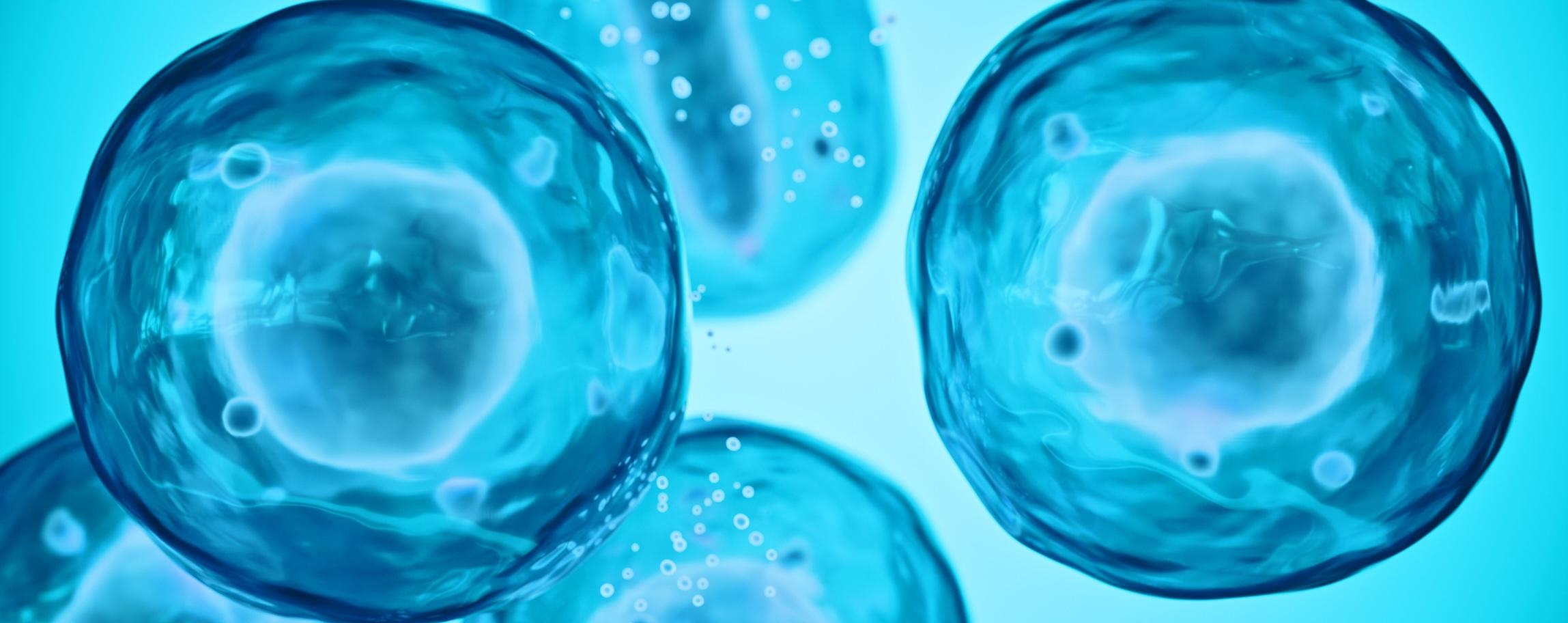
In the past two decades, organoids have transformed from a conceptual curiosity into one of the most powerful tools in modern biomedical research. These self-organizing, three-dimensional (3D) cellular structures recreate key features of human organs in vitro, providing an unprecedented bridge between reductionist two-dimensional (2D) culture systems and the physiological complexity of living tissue. Derived from embryonic, induced pluripotent, or adult stem cells, organoids can replicate essential aspects of organ architecture, cellular diversity, and function — effectively serving as “mini-organs” in a dish.
The field’s roots trace to early efforts in the 1980s and 1990s to grow epithelial stem cell aggregates, but researchers were only able to get organoids to begin to self-organize and persist long-term with the advent of refined stem-cell protocols and extracellular matrices.1 These advances allowed researchers to recapitulate the morphogenetic cues and microenvironmental signals that drive tissue organization in vivo, enabling the formation of organ-like domains, such as intestinal crypts, cortical layers, or hepatic lobules. What began as a developmental biology breakthrough rapidly became a transformative platform for translational science.
The appeal of organoids lies not only in their physiological fidelity but in their ability to address the long-standing translational bottleneck between animal models and human outcomes. Despite their utility, conventional 2D cultures fail to capture multicellular interactions, and animal models often exhibit poor predictive value for human drug responses.2,3 Organoids offer a more accurate, ethically preferable, and scalable means to study disease, screen compounds, and predict therapeutic efficacy. They retain patient-specific genetics and phenotypes, allowing investigators to model individual disease trajectories and drug sensitivities with remarkable precision.
Today, organoids are being integrated across the pharmaceutical and biomedical landscape, from disease modeling and personalized drug discovery to toxicology testing and regenerative medicine.4 Industrial players are scaling production, standardizing protocols, and automating analysis pipelines, signaling a transition from academic innovation to applied technology. As these miniature organ systems continue to mature, they are reshaping the foundation of preclinical research and bringing the promise of truly human-based science within reach.
From Stem Cells to Self-Organization: Building the Mini-Organ
At its core, an organoid is a self-organizing 3D construct derived from stem cells that recapitulates the architecture and function of its tissue of origin. What distinguishes an organoid from a spheroid or simple multicellular aggregate is not just its shape or dimensionality but its developmental logic: the ability to autonomously organize into hierarchically structured, functionally specialized regions through the same cues that guide organogenesis in vivo.1,5
Organoids can arise from several cellular sources, each contributing unique advantages and constraints. Embryonic stem cells and induced pluripotent stem cells (iPSCs) offer pluripotency, enabling differentiation into nearly any tissue type and the modeling of early developmental processes.5 Adult stem cells, typically harvested from epithelial tissues, such as the intestine, liver, or airway, provide lineage-restricted yet physiologically mature organoids ideal for studying homeostasis, regeneration, or disease mechanisms. Tumor-derived organoids extend this concept to oncology, preserving the intratumoral heterogeneity, genetic mutations, and microenvironmental interactions that drive individual patient responses to therapy.6 This cellular versatility underpins the broad applicability of organoids across research domains.
Central to organoid formation is the principle of self-organization, a process in which stem cells respond to intrinsic gene regulatory networks and extrinsic biochemical cues to recreate their native microenvironment, or niche. Within an appropriately supportive matrix, these cells spontaneously establish polarity, patterning, and compartmentalization, forming crypt-villus structures in intestinal models, nephron-like tubules in kidney organoids, or layered neuronal tissues in cortical organoids.1 The orchestrated choreography of signaling pathways, such as Wnt, BMP, Notch, and FGF, recapitulates embryonic tissue specification, while cell–cell and cell–matrix interactions provide the mechanical and biochemical context needed for spatial organization.
The extracellular matrix (ECM) serves as both the scaffold and the signaling environment guiding this morphogenesis. Most organoids historically rely on Matrigel, a gelatinous mixture derived from mouse sarcoma rich in laminin, collagen IV, and entactin. Although Matrigel supports robust growth, its undefined composition, lot-to-lot variability, and non-human origin limit standardization and clinical translation. This has prompted a surge in efforts to engineer more consistent and physiologically relevant alternatives, such as decellularized ECM (dECM) hydrogels derived from human tissues and synthetic matrices with tunable stiffness and ligand presentation.7,8 These next-generation scaffolds allow tighter control over mechanical and biochemical parameters, enabling improved reproducibility, organ-specific maturation, and compatibility with scalable manufacturing.
Despite these advances, organoid cultures remain inherently variable. Differences in starting cell populations, growth factors, and matrix composition can yield heterogeneous outcomes even within the same protocol. Batch-to-batch variability and incomplete maturation, especially in vascularization, immune competence, and metabolic function, continue to challenge reproducibility and physiological relevance.9 Researchers are increasingly integrating microfluidic perfusion, biomaterial engineering, and defined culture media to mitigate these limitations, but the field remains at a transition point between artisanal and standardized science.
At its most fundamental level, however, the recipe for organoid formation has changed little: self-renewing stem cells, precise differentiation cues, a supportive extracellular matrix, and time. Together, these ingredients allow human cells to recapitulate the organizing principles of life itself: building from the bottom up, guided by their intrinsic blueprint rather than external design.
Engineering the Environment: Matrices, Mechanics, and Microfluidics
As organoid systems have grown more sophisticated, the field’s attention has shifted from cellular differentiation to the engineering of the microenvironment that sustains it. The physical and biochemical context in which cells grow is now recognized as just as important as the cells themselves, dictating morphology, maturation, and function. To make organoids more physiologically relevant — and more reproducible — researchers have turned to innovations in materials science, bioreactor design, and microengineering that move the culture environment closer to the dynamic conditions of living tissue.
One of the most significant advances has been the move away from Matrigel toward defined and decellularized ECM (dECM) hydrogels that better mimic native tissue composition. dECM matrices derived from human sources preserve the structural proteins, glycosaminoglycans, and bioactive motifs that guide cell adhesion and differentiation while eliminating the undefined variability inherent in tumor-derived substrates.7 Because these hydrogels can be customized for stiffness, porosity, and biochemical composition, they enable precise control over cell–matrix interactions and mechanical signaling. This tunability has allowed researchers to reproduce tissue-specific microenvironments (e.g., soft matrices for neural differentiation, stiffer substrates for intestinal or hepatic lineages) thereby improving both the fidelity and scalability of organoid culture.
Beyond static scaffolds, microfluidic systems have introduced a new level of physiological realism. By integrating perfusable channels, these “organ-on-chip” platforms recreate the continuous nutrient flow, shear stress, and waste removal that characterize in vivo tissues.8,9 Dynamic perfusion not only improves metabolic stability and size scalability but also promotes structural maturation, including the development of vascular-like networks within the organoid mass. Advances in 3D bioprinting have extended this progress further, allowing precise spatial deposition of multiple cell types and ECM materials in architectures that mirror native tissues. Such additive manufacturing approaches support the creation of patterned microvasculature and the assembly of multi-organoid constructs — so-called “assembloids” — capable of modeling inter-organ communication.
Parallel progress has been made in sensorized bioreactors and real-time monitoring technologies, which bring process control and analytical rigor to organoid culture. Embedded oxygen, pH, and metabolite sensors allow continuous feedback on culture conditions, supporting automated adjustments to maintain homeostasis. Integration with machine vision enables non-invasive tracking of morphology, growth kinetics, and functional activity, creating closed-loop systems that blur the line between biological and engineered intelligence. These platforms not only improve reproducibility but also generate high-content data sets linking microenvironmental parameters to phenotypic outcomes, which are critical for both research standardization and regulatory acceptance.
Perhaps the most exciting step in the evolution of organoid platforms has been their integration with organ-on-chip and microphysiological systems, enabling complex, multi-tissue modeling. By linking organoids through microfluidic channels, researchers can now study pharmacokinetics and organ–organ interactions in controlled human-relevant systems.9,10 Liver and intestinal organoids can be connected to mimic first-pass metabolism, while cardiac and vascular organoids can be coupled to model drug-induced cardiotoxicity. These hybrid systems promise a more predictive framework for drug development that captures systemic physiology without the ethical and translational drawbacks of animal models.
Meanwhile, the frontier continues to expand beyond structural fidelity toward functional completeness. Efforts to generate immune-competent organoids are bringing elements of adaptive and innate immunity into play, facilitating studies of inflammation, infection, and immunotherapy response.10 Similarly, innervated organoids, particularly in gut and brain models, introduce neural circuitry that enables electrophysiological readouts and dynamic behavioral responses. Together, these innovations suggest a trajectory toward organoids that are not only anatomically and biochemically correct but also physiologically alive in ways once thought impossible.
What emerges is a convergence of disciplines — cell biology, materials science, bioengineering, and data analytics — transforming organoid systems from fragile lab curiosities into robust, industrially scalable technologies. By engineering the environment as carefully as the biology, scientists are learning to let human cells express their full potential, forming living systems that increasingly resemble the organs they aim to model.
Organoids as Models of Human Disease
The greatest validation of organoid technology lies in its ability to replicate the complexity of human disease more faithfully than any other in vitro system. By capturing the genetic, structural, and functional characteristics of native tissues, organoids provide a versatile platform to study both inherited and acquired disorders across organ systems. Whether derived from patient samples or reprogrammed iPSCs or engineered through CRISPR-based modification, they enable scientists to model disease progression, dissect molecular mechanisms, and test therapeutic responses within a physiologically relevant human context.
Genetic and Developmental Disorders
The earliest demonstrations of organoid utility came from their application to rare monogenic diseases, where patient-derived or gene-edited lines offered a direct link between genotype and phenotype. Intestinal organoids from cystic fibrosis patients, for example, reproduce the characteristic ion transport defects caused by CFTR mutations and have been used to test modulator therapies that restore channel function.5 Similarly, retinal and cerebral organoids derived from iPSCs allow researchers to trace the consequences of developmental mutations on tissue formation, providing windows into conditions such as microcephaly or retinitis pigmentosa.6
The ability to introduce or correct mutations through CRISPR–Cas9 gene editing has further expanded the precision of these models. Researchers can now generate isogenic control lines that differ only at a single pathogenic locus, eliminating background variability and enabling clear causal inferences. These systems not only accelerate mechanistic discovery but also serve as preclinical testbeds for gene and cell therapies, allowing safety and efficacy assessments in a human context long before clinical translation. For diseases where animal models fail to capture human-specific developmental cues, such as certain congenital heart or neural tube defects, organoids offer a uniquely faithful representation of early organogenesis and tissue morphodynamics.
Infectious Diseases
Organoids have also become indispensable tools for understanding host–pathogen interactions, particularly in epithelial tissues that form natural barriers to infection. Gut, lung, and airway organoids replicate the architecture, receptor profiles, and mucus-secreting physiology of these interfaces, making them ideal for studying microbial invasion and immune signaling. Gastrointestinal organoids, for example, have been used to model infections with rotavirus, norovirus, and Helicobacter pylori, revealing how pathogens manipulate epithelial polarity and disrupt barrier function.11 Airway organoids, meanwhile, provided critical insight during the COVID-19 pandemic by demonstrating the specific cellular tropism of SARS-CoV-2 and the downstream inflammatory cascades it triggers.
These models offer unprecedented resolution for dissecting the molecular dialogue between host and pathogen, including interferon signaling, epithelial regeneration, and microbiome–immune crosstalk. Importantly, because organoids can be derived from different donors, they also capture population-level variability in susceptibility and response, enabling studies of genetic or epigenetic determinants of infection outcomes. Such insights are guiding the development of antiviral drugs and vaccines while minimizing reliance on animal systems that poorly mimic human pathogen dynamics.
Cancer and Precision Oncology
Perhaps no field has embraced organoid technology more rapidly than oncology. Patient-derived organoids (PDOs) grown directly from surgical or biopsy specimens preserve the heterogeneity of primary tumors, including driver mutations, epigenetic landscapes, and microarchitectural features, often lost in conventional cell lines. These models provide a powerful bridge between molecular profiling and clinical decision-making.12
In drug discovery, large biobanks of PDOs spanning colorectal, breast, pancreatic, and lung cancers are being used to screen compound libraries and identify genotype–drug response correlations. Because PDOs maintain patient-specific mutational signatures and drug resistance mechanisms, they serve as predictive avatars for precision oncology trials.2 Studies comparing in vitro organoid responses with clinical outcomes have shown striking concordance, suggesting that these systems could soon guide real-time therapeutic choices.
PDOs are also revolutionizing our understanding of tumor evolution and microenvironmental interactions. When co-cultured with fibroblasts, immune cells, or vasculature-mimicking endothelium, they reveal how cancer cells adapt to or exploit their surroundings. Combined with CRISPR-based perturbation screening and single-cell sequencing, organoid platforms now enable functional annotation of oncogenic variants and the mapping of drug resistance trajectories with unparalleled detail. For pharmaceutical developers, they offer both a more predictive preclinical model and an ethically sound replacement for large-scale animal testing.
Neurodegeneration and Beyond
In the neurosciences, organoids have unlocked experimental frontiers previously out of reach. Brain organoids derived from human iPSCs reproduce many of the cellular layers and signaling circuits of the developing cortex, allowing mechanistic studies of neurodevelopmental and neurodegenerative disease alike. Models of Alzheimer’s and Parkinson’s disease generated from patient lines recapitulate hallmark features, such as amyloid-β accumulation, tau pathology, and dopaminergic neuron loss, providing a living system in which to test candidate therapeutics and observe long-term neuronal dysfunction.9 Retinal organoids have proven similarly valuable for investigating photoreceptor degeneration and for screening gene- or stem cell—based interventions aimed at vision restoration.13
Beyond pathology, neuronal organoids are beginning to illuminate how genetic and environmental factors intersect in conditions such as autism spectrum disorder and schizophrenia, revealing network-level alterations in connectivity and synaptic activity. As researchers integrate microglia, vascular elements, and sensory inputs, these constructs are inching closer to fully functional models of human brain physiology. The ability to monitor electrophysiological activity and synaptic plasticity in vitro opens possibilities not just for disease modeling but for the exploration of human cognition and drug effects on neural circuitry.
Across all these domains, organoids achieve a level of fidelity unmatched by earlier model systems, aligning with human tissue at genomic, histologic, and functional levels.1 Yet the question “how close is close enough?” remains an active one. While no in vitro system perfectly replicates the complexity of an intact organ, organoids now provide a practical and ethically defensible means to study disease mechanisms in a setting that reflects the diversity and specificity of human biology. As their precision improves, they promise not only to complement but, in many cases, to replace traditional animal models as the gold standard for translational research.
Organoids in Drug Development and Screening
In the pharmaceutical industry’s relentless pursuit of better predictivity, organoids have emerged as one of the most transformative technologies in preclinical R&D. Their ability to replicate the cellular diversity, architecture, and genetic fidelity of human tissues gives them a unique position in the translational pipeline, bridging the gap between basic biology and clinical reality. Once confined to academic laboratories, organoid systems are now being integrated into discovery, safety, and efficacy workflows across the drug development continuum, helping companies design smarter experiments, reduce attrition, and accelerate time to market.
At the foundation of this transformation is the use of organoid panels as predictive preclinical tools. Unlike traditional immortalized cell lines that represent a single genotype or tumor subtype, panels of patient-derived organoids capture the genomic and phenotypic heterogeneity that defines real-world disease.3,14 These panels enable population-scale screening of therapeutic candidates across diverse genetic backgrounds, generating a rich matrix of efficacy and toxicity data that more accurately forecasts clinical response distributions. Pharmaceutical developers are increasingly deploying such panels to prioritize compounds for development, de-risk clinical trials, and identify biomarkers of response or resistance. Because organoids preserve patient-specific mutations and gene expression profiles, they also serve as precision avatars for testing targeted or combination therapies before first-in-human studies.
Organoids consistently outperform 2D cultures and animal models in both reproducibility and predictivity. Two-dimensional systems lack the multicellular organization and gradients of oxygen, nutrients, and metabolites that shape real tissue physiology, while animal models often fail to capture human-specific signaling pathways or immune responses. In contrast, organoids reproduce many of the microenvironmental interactions — cell–cell, cell–matrix, and paracrine —that determine therapeutic outcomes in vivo.14 They generate dose–response curves, metabolic profiles, and cytotoxicity signatures that align closely with clinical data, offering early insights into efficacy and potential adverse effects. Their modular nature further allows scientists to integrate key elements of tissue physiology, such as vasculature, innervation, and immune components, at will, customizing each model to the therapeutic mechanism of interest.
As organoids move deeper into the drug development workflow, they are being incorporated into multi-parameter assay systems that simultaneously measure efficacy, toxicity, and pharmacokinetic behavior. Organoids now serve as testbeds for absorption, distribution, metabolism, and excretion (ADME) modeling, enabling compound optimization before costly in vivo studies.2,11 For instance, liver organoids provide predictive data on cytochrome P450 activity and metabolite generation, while intestinal and kidney organoids shed light on permeability and clearance. Because they can be co-cultured with endothelial or immune cells, organoids also allow integrated evaluation of inflammation, fibrosis, or immune-mediated toxicities, endpoints that have historically been difficult to model preclinically.
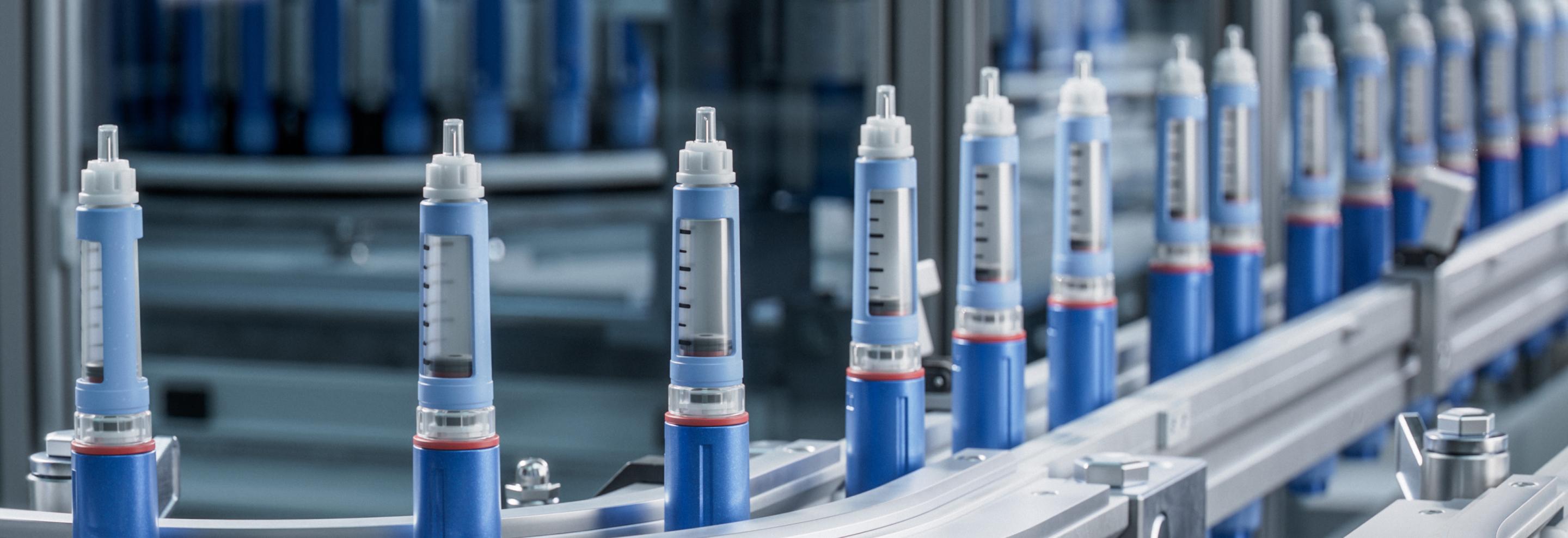
The field’s maturation has been propelled by advances in automation and high-throughput screening. Early organoid protocols were labor-intensive and artisanal, limiting their scalability for pharmaceutical applications. That bottleneck is rapidly disappearing. Robotic liquid-handling systems, standardized microplate formats, and automated imaging platforms now support thousands of parallel organoid assays, while artificial intelligence (AI)–assisted image analysis quantifies morphological and functional responses with single-cell precision.6,15 These platforms can measure proliferation, apoptosis, metabolic flux, and even contractile or electrophysiological behavior depending on tissue type. Coupled with machine learning, the resulting data sets enable predictive modeling of therapeutic response and safety liabilities across hundreds of compounds simultaneously.
The combination of high-throughput capability and patient relevance has fueled the creation of organoid biobanks and matched screening programs. Large academic and commercial initiatives now maintain libraries of organoids derived from diverse tissues, diseases, and patient populations.12 These collections enable cross-comparison of drug sensitivities and support population-level studies of pharmacogenomic variation. In oncology, biobanks of PDOs are being linked to clinical registries, allowing investigators to correlate in vitro drug sensitivity with real patient outcomes. In other disease areas, such as cystic fibrosis and inflammatory bowel disease, organoid assays are beginning to inform therapeutic selection on a patient-by-patient basis, offering a glimpse of the future of precision medicine.
As adoption accelerates, regulators are also beginning to acknowledge the value of organoid systems in preclinical evaluation. Under the FDA Modernization Act 2.0 and parallel initiatives in Europe, organoids and other “new approach methodologies” (NAMs) are gaining traction as acceptable alternatives to certain animal studies, provided they demonstrate validated performance and reproducibility.3 Regulatory convergence around NAMs marks a pivotal shift in the drug development paradigm: moving away from animal-based safety surrogates toward human-relevant platforms. Organoid models, particularly when combined with microphysiological systems and computational modeling, are likely to play a central role in this evolution.
Still, challenges remain before organoids can be fully standardized as regulatory tools. Reproducibility across laboratories, batch variation in matrices, and the need for robust reference data are persistent issues. Yet the trajectory is clear: as protocols become more defined and analytics more automated, organoids are transitioning from exploratory models to validated, auditable components of the pharmaceutical quality ecosystem.
The practical implications for industry are profound. By integrating organoids into early screening, companies can eliminate non-predictive leads sooner, focus resources on compounds with higher translational potential, and uncover liabilities before they derail clinical trials. Beyond economics, the ethical and scientific advantages are equally compelling, replacing large-scale animal testing with systems that reflect the molecular and cellular complexity of the human body. In this sense, organoids represent not just a technical innovation but a philosophical one, redefining what it means to test drugs on humans, without ever entering a human body.
As the industry continues to align around this paradigm, organoids stand poised to reshape every phase of the drug development pipeline, from discovery and optimization to safety testing and precision therapy design. Their growing predictive power is already accelerating the feedback loop between laboratory insight and clinical impact, bringing pharmaceutical R&D closer to its long-standing goal of translating biology into better medicine, faster and more humanely than ever before.
Industrialization and Standardization
For organoids to fulfill their promise as the foundation of next-generation biomedical research and drug development, they must evolve from handcrafted biological experiments into industrially reproducible products. Academic ingenuity has shown what organoids can do; the challenge now is ensuring that every batch, across every lab or biomanufacturing facility, performs the same way. This transition from bespoke science to scalable technology is defining the next phase of organoid innovation, bringing with it the rigor of process control, data integrity, and quality assurance long familiar in pharmaceutical manufacturing.
Efforts to establish standardized culture conditions and quality control (QC) parameters are central to this evolution. Early organoid research was highly artisanal, with subtle differences in stem-cell source, matrix formulation, and growth factor timing leading to dramatic variability in morphology and function. Initiatives are now converging around defined media formulations, quantifiable maturity markers, and standardized endpoints for organoid quality and performance.1,9 These metrics — covering morphology, viability, gene expression, and functional outputs — are becoming the foundation of reproducible biomanufacturing workflows. At the same time, interlaboratory consortia and reference databases are helping to harmonize protocols across research centers, ensuring that organoids produced in different settings can be directly compared or pooled into shared biobanks.
Commercial suppliers have accelerated this process by offering assay-ready organoids that meet predefined specifications for purity, differentiation stage, and response predictability.4,15 These standardized products, available for a variety of tissues and disease indications, remove much of the variability and lead time associated with in-house derivation. They allow pharmaceutical companies to focus on experimental design and data interpretation rather than cell-culture optimization, bringing organoids into the same operational category as other validated biological reagents. In parallel, contract research organizations and biotech startups are providing end-to-end organoid assay services, complete with QC certification, data analysis pipelines, and regulatory documentation, to support the growing demand for reproducible, auditable preclinical testing.
As scale and throughput increase, automation, robotics, and AI are becoming indispensable tools for maintaining consistency and interpretability. Robotic liquid handlers can seed, feed, and image thousands of organoids with precision, eliminating human variability while supporting high-throughput experimentation. Automated imaging systems coupled with AI-driven morphology scoring provide objective, quantitative assessments of organoid health, differentiation, and treatment response.4,8 These platforms not only improve reproducibility but also generate vast data sets that feed machine-learning algorithms, uncovering subtle phenotypic correlations and predictive biomarkers that would be invisible through manual inspection. The result is a closed-loop ecosystem in which automation ensures consistency, and AI continuously refines understanding.
The parallels with biomanufacturing are striking. As organoids transition from exploratory models to regulated research and therapeutic tools, principles such as batch tracking, material traceability, and data integrity are becoming nonnegotiable. Each culture can be treated as a “lot” with its own provenance, digital record, and performance profile, mirroring the documentation frameworks used in GMP production. Analytical methods adapted from process analytics and quality-by-design (QbD) paradigms are now being applied to organoid generation, allowing process deviations to be detected and corrected in real time. This industrial mindset aligns naturally with the regulatory and operational standards that pharmaceutical companies already uphold, positioning organoids for seamless integration into established development pipelines.
Yet industrialization also brings ethical and intellectual property (IP) complexities, especially for organoids derived from patient material. Questions around ownership, consent, and secondary use of biospecimens are intensifying as biobanks expand and commercial applications multiply.1 Unlike anonymized cell lines, patient-derived organoids carry genetic fingerprints that may be linked back to individuals, requiring stringent governance for data privacy and access control. Moreover, as companies patent specific derivation protocols or organoid compositions, tensions are emerging between open scientific collaboration and proprietary protection. Balancing transparency with commercial viability will be critical to sustaining both innovation and public trust.
The QC challenge that underlies all of these efforts is as much philosophical as technical. Reproducibility in living systems demands both standardization and flexibility: the ability to define consistent processes while accommodating biological variability that reflects real human diversity. Achieving that balance will determine how far organoids can advance as a regulated, industrial platform.
As these systems mature, the boundaries between research biology and manufacturing science are dissolving. Organoids are no longer one-off creations assembled by expert hands; they are evolving into reliable, traceable, and data-rich tools that can be mass-produced without sacrificing fidelity. By embedding the rigor of quality systems and the intelligence of automation into their production, the life sciences industry is ensuring that organoids can scale with confidence, turning biological complexity into a controlled, reproducible resource for discovery and development at industrial speed.
The Next Frontier: Regeneration, Integration, and Intelligence
As organoid technology matures, its potential is expanding beyond disease modeling and drug screening into realms that could fundamentally reshape medicine itself. The next frontier is not simply about reproducing organs in miniature, but about using organoids as building blocks for regeneration, system-level integration, and even biointelligence. What began as an experimental platform for understanding development is now converging with tissue engineering, computational biology, and AI to redefine the boundaries of what living systems can do.
In regenerative medicine, organoids are emerging as scalable, patient-specific units for tissue repair and replacement. Their ability to self-organize, differentiate, and maintain long-term viability makes them ideal candidates for reconstructing damaged or diseased tissues. Liver and intestinal organoids, for instance, have already been transplanted into animal models, where they integrate with host vasculature and restore partial function.7,13 dECM scaffolds and 3D bioprinting are enabling these organoids to be positioned precisely within damaged tissues, enhancing engraftment and promoting regeneration. In the longer term, combining organoids with vascular grafts, immune modulation, and biomaterial interfaces could yield implantable constructs that replace or augment failing organs. While ethical and safety barriers remain before widespread clinical use, early successes suggest a plausible route toward autologous tissue replacement — personalized regeneration built from the patient’s own cells.
Beyond the single organ, researchers are beginning to explore organoid–organ communication through multi-organoid assemblies known as assembloids. These constructs link organoids representing different tissues (e.g., brain–retina, gut–liver, or pancreas–islet complexes) to study inter-organ signaling, metabolism, and systemic disease processes.9 By connecting organoids through microfluidic circuits or shared vasculature, scientists can simulate the integrated physiology of the human body on a miniature scale. This approach is not only advancing drug toxicity testing and metabolic modeling but also offering unprecedented insight into how organs co-regulate each other in health and disease. In parallel, computational modeling is enabling the creation of digital twins — virtual counterparts of organoid systems that mirror their structural and functional dynamics in silico. Integrating organoid data streams with digital twin platforms allows researchers to simulate therapeutic interventions, predict outcomes, and refine protocols with reduced experimental burden.
Perhaps the most visionary step forward lies in the concept of organoid intelligence. Brain organoids, with their layered neuronal structures and spontaneous electrical activity, have already demonstrated primitive learning-like behaviors and signal processing capabilities.9 This emerging field — sometimes described as neurobiocomputing — proposes that living neuronal networks could eventually perform adaptive computations that complement or even surpass silicon-based AI in energy efficiency and pattern recognition. While still in its infancy, organoid intelligence invites profound questions about the intersection of biology and information science: how living systems encode, process, and remember information, and whether these capabilities can be harnessed for biomedical or computational applications.
At the same time, AI is returning the favor by enhancing organoid science itself. Machine learning algorithms are increasingly used to optimize differentiation protocols, predict culture outcomes, and analyze high-content imaging data.4 By correlating experimental variables with organoid morphology, function, and molecular signatures, AI tools can identify subtle relationships invisible to human observers, accelerating discovery and improving reproducibility. As data sets from organoid biobanks, screening assays, and microphysiological systems expand, these computational frameworks will become essential for modeling complex biology, designing personalized interventions, and guiding automated manufacturing.
Taken together, these advances represent a profound synthesis of biology and technology. Organoids are no longer passive models of human physiology; they are active participants in the creation of new therapeutic and computational paradigms. Whether as implantable tissues, integrated system models, or living processors, organoids are pushing the life sciences toward a future where regeneration, prediction, and intelligence converge.
Conclusion: Toward Predictive, Ethical, and Human-Centered Preclinical Science
In just two decades, organoids have progressed from experimental curiosities to essential instruments of translational medicine. What began as an ambitious attempt to coax stem cells into forming miniature organs has matured into a robust scientific and industrial framework that is reshaping the drug development process.1 Organoids now stand alongside other transformative technologies, such as induced pluripotent stem cells, CRISPR editing, and organ-on-chip systems, as pillars of a more human-centered approach to biomedical innovation.
Their impact extends across every layer of research and development. Organoids are replacing or refining animal models, capturing human tissue complexity in ways that dramatically improve translational accuracy. They have already increased predictive success rates in early-phase testing, reduced reliance on animal experimentation, and accelerated the feedback loop between discovery and clinical validation. In parallel, patient-derived organoid platforms are ushering in the era of precision medicine, where individual genetics and disease phenotypes directly inform therapeutic strategy. The vision of truly personalized preclinical testing — one organoid per patient — is moving from concept to operational reality.
Yet, as the field moves toward industrial maturity, several challenges remain. Cost and complexity continue to limit scalability, while inconsistencies in differentiation protocols and culture matrices hinder standardization. Most organoids still lack the full physiological integration — vascular, immune, or endocrine — that defines functional organs. The community’s next task will be to balance biological fidelity with manufacturability, ensuring that organoids can serve as both scientifically rigorous and operationally feasible tools across the pharmaceutical value chain.
Despite these hurdles, the trajectory is unmistakable. Organoids have already become the most faithful in vitro representation of human biology available, and their influence is expanding far beyond the laboratory bench. As regulatory bodies embrace new approach methodologies and as automation brings reproducibility within reach, organoids are set to become the connective tissue between experimental biology and therapeutic discovery.
References
1. Trentesaux, Coralie, et al. “Harnessing synthetic biology to engineer organoids and tissues.” Cell Stem Cell. 30: 10–19 (2023).
2. Gopallawa, Indiwari, et al. “Applications of Organoids in Advancing Drug Discovery and Development.” Journal of Phamaceutical Sciences. 113: 2659–2667 (2024).
3. Kelder, Regina. “Organoids in Drug Development: Evolving and Expanding.” Eureka. 24 Apr. 2024.
4. Srinivasan, Maithreyan. “2025 Trends: Organoids.” Genetic Engineering & Biotechnology News. 6 Jan 2025.
5. Yang, Siqi, et al. “Organoids: The current status and biomedical applications.” MedComm (2020). 4: e274 (2020).
6. Candana, J Jeya, et al. “Human pluripotent-stem-cell-derived organoids for drug discovery and evaluation.” Cell Stem Cell. 30: P571–591 (2023).
7. Li, Chen, et al. “Enhancing organoid culture: harnessing the potential of decellularized extracellular matrix hydrogels for mimicking microenvironments.” Journal of Biomedical Science. 31: 96 (2024).
8. Maffeis, Vaviana, et al. “Synthetic Cells Revisited: Artificial Cell Construction Using Polymeric Building Blocks.” Advanced Science. 11: 2305837 (2023).
9. Han, Xinxin, et al. “Landscape of human organoids: Ideal model in clinics and research.” The Innovation. 5: 100620 (2024).
10. Goldrick, Caoimhe, et al. “3D multicellular systems in disease modelling: From organoids to organ-on-chip.” Front. Cell Dev. Biol. Sec. Stem Cell Research. 1 Feb. 2023.
11. Zhou, Li, et al. “Gastrointestinal tract organoids as novel tools in drug discovery.” Front. Pharmacol. Sec. Experimental Pharmacology and Drug Discovery. 29 Aug. 2024.
12. Yao, Qigu, et al. “Organoids: development and applications in disease models, drug discovery, precision medicine, and regenerative medicine.” MedComm (2020). 5: e735 (2024).
13. Makesh, Karnika Y, et al. “A Concise Review of Organoid Tissue Engineering: Regenerative Applications and Precision Medicine.” Organoids. 4: 16 (2025).
14. Putker, Marrit. “Why Organoids Are Superior for Drug Screening: Answering Top Research Questions.” Crown Bioscience. 27 May 2024.
15. Armstrong, Reece. “Pushing drug discovery with stem cells, organoids and 3D cell cultures.” DDW Online. 16 Apr. 2025.